
Table of contents
- Technology Adoption in Pune Neurosurgery 2025-2026
- Neuronavigation: The Technology That Made MISS Possible
- Intraoperative MRI: Seeing the Brain During Surgery
- Robotic Spine Surgery: Precision Implant Placement
- 5-ALA Fluorescence-Guided Surgery for Brain Tumors
- Intraoperative Neurophysiological Monitoring (IONM)
- AI-Assisted Diagnosis and Surgical Planning
- What This Means for Patients in Pune and PCMC
- Frequently Asked Questions
KEY TAKEAWAYS
- Neuronavigation the brain surgeon’s GPS is now standard at hospitals in Pune’s Baner-Wakad corridor, enabling sub-millimetre surgical precision for brain tumor and skull base surgery.
- Intraoperative MRI (iMRI), available at select Pune centres, allows surgeons to confirm tumor resection completeness while the patient is still on the operating table increasing gross total resection rates by 20 to 30% for gliomas.
- Robotic spine surgery systems (such as Mazor X and Globus ExcelsiusGPS) guide pedicle screw placement with 1 to 2 mm accuracy significantly reducing radiation exposure for both patient and surgical team.
- AI-assisted MRI analysis is increasingly used for tumor boundary delineation, surgical planning and intraoperative guidance reducing reliance on surgeon judgement alone for critical anatomical decisions.
- 5-ALA fluorescence-guided surgery makes malignant glioma cells glow pink under blue-violet light helping surgeons remove more tumor while protecting healthy brain, improving progression-free survival.
- None of these technologies replace the neurosurgeon’s judgement and technical skill they amplify it, providing real-time data that informs decisions that remain fundamentally human.
- Dr. Sarang Gotecha uses neuronavigation, operating microscopy and IONM as standard at his Pune neurosurgery practice ensuring patients across Baner, Wakad and PCMC access technology-enhanced surgery locally.
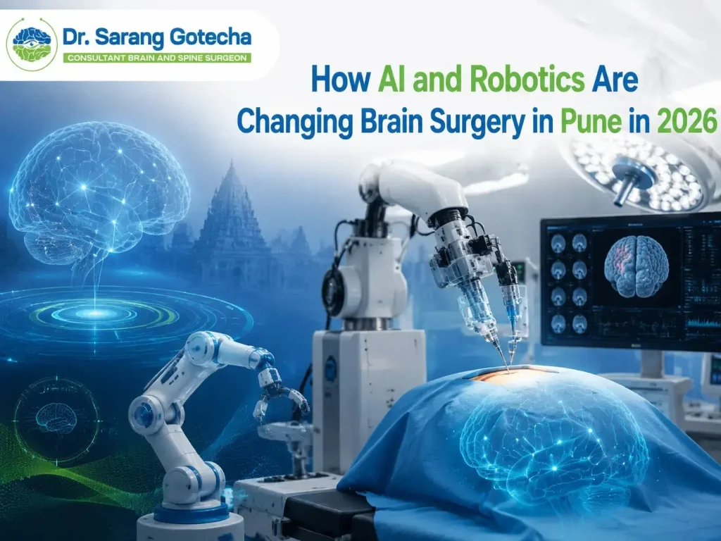
The image of brain surgery that most Pune families carry a surgeon peering into an open skull, relying entirely on experience and instinct is outdated. Modern neurosurgery in 2026 is a technology-rich discipline where GPS-precision navigation, real-time MRI imaging, robot-guided implant placement and AI-assisted diagnostics work alongside the surgeon’s hands and judgement to produce safer, more precise outcomes.
This guide explains the key technologies transforming neurosurgery in Pune, what they do, which are available locally and what they mean for patients and families choosing where and with whom to have brain or spine surgery.
QUICK FACTS
Neuronavigation Accuracy: Under 1 mm sub-millimetre 3D localisation
iMRI GTR Rate Improvement for GBM: Increases gross total resection from ~36% to ~65%
Robotic Pedicle Screw Accuracy: Over 95% within 2 mm of planned positio
5-ALA Fluorescence Surgery: Improves complete GBM resection from 36% to 65%
IONM Neurological Deficit Reduction: Reduces permanent deficits by 50 to 75% for eloquent area surgery
Availability in Pune: Neuronavigation and IONM standard at Baner corridor hospitals
Technology Adoption in Pune Neurosurgery 2025-2026
| Technology | Availability in Pune | Benefit | Used By Dr. Sarang Gotecha |
| Neuronavigation | Standard most neurosurgical centres | Sub-mm targeting, MISS enables | Yes |
| Operating Microscope | Universal | Magnification, illumination | Yes |
| Intraoperative Neurophysiology (IONM) | Available select centres | Real-time function monitoring | Yes |
| 5-ALA Fluorescence Surgery | Available select Pune centres | GBM resection improvement | Yes (for GBM cases) |
| Intraoperative MRI (iMRI) | Available 1 to 2 Pune centres | Resection completeness confirmation | Available via referral |
| Robotic Spine Surgery | Available increasing adoption Pune | Screw placement accuracy | Available via referral |
| AI Diagnostic Assistance | Emerging selected tools | Tumor segmentation, planning | Available tools used |
| Awake Craniotomy | Available select centres | Eloquent area function mapping | Yes |
Neuronavigation: The Technology That Made MISS Possible
Neuronavigation is the most widely adopted and impactful surgical technology in modern neurosurgery. Its adoption has been described as the single most important technical advance in brain surgery since the operating microscope. Understanding how it works demystifies much of what feels mysterious about modern brain surgery.
Before surgery, the patient’s MRI is loaded into the navigation workstation. The workstation creates a three-dimensional digital model of the patient’s brain, showing the tumor, critical structures, vessels and ventricles in precise anatomical relationship. In the operating room, reference arrays are fixed to the patient’s head and registered to the MRI creating a live correspondence between the physical patient and the digital model.
From this point, every tracked surgical instrument shows its position on the 3D brain model in real time. The surgeon can plan the optimal trajectory to the tumor the shortest, safest path avoiding eloquent cortex, major vessels and venous structures before making the first incision. Mini craniotomy (3 to 4 cm bone flap) and keyhole approaches become reliable rather than risky because the navigation system confirms that the small opening is precisely positioned.
Neuronavigation is now standard at hospitals in Pune’s Baner-Wakad corridor. Its availability has ended the argument for travelling to Mumbai for brain tumor surgery that requires precise targeting — the technology gap that once justified that journey no longer exists for most standard and intermediate-complexity cases.
Intraoperative MRI: Seeing the Brain During Surgery
The standard approach to assessing surgical completeness is a post-operative MRI performed 24 to 48 hours after surgery. By then, the patient has been closed, transferred to ICU, woken from anaesthesia and transported to the MRI unit. If significant residual tumor is found on the post-operative scan, a second surgery is required to complete the resection with all the additional risks of repeat craniotomy.
Intraoperative MRI eliminates this problem by bringing the MRI scanner into the operating room. At Pune centres equipped with iMRI, the surgeon can pause the resection, scan the patient without moving them from the operating table, review the images and if residual tumor is identified continue the resection immediately before closing the wound.
The clinical impact is significant. For glioblastoma surgery, iMRI increases gross total resection (complete removal of the contrast-enhancing tumor) rates from approximately 36% with standard surgery to 65% an increase directly associated with improved median survival. For pituitary adenomas, iMRI confirms complete sellar clearance before the nasal endoscope is withdrawn. For gliomas near eloquent cortex, iMRI combined with awake craniotomy provides the most comprehensive information available for maximising safe resection.
Robotic Spine Surgery: Precision Implant Placement
Pedicle screw placement for spinal fusion whether for fracture fixation, spondylolisthesis, degenerative disc disease or spinal tumor has traditionally relied on the surgeon’s anatomical knowledge and fluoroscopic X-ray guidance. Correct screw position within the narrow pedicle (the bony bridge connecting the vertebral body to the posterior arch) requires precise trajectory a misplaced screw can injure adjacent nerve roots or the spinal cord.
Robotic spine surgery systems (including the Mazor X Stealth and Globus ExcelsiusGPS, with increasing adoption at Pune’s surgical centres) use pre-operative CT-based planning and robotic arm guidance to position screw guides with sub-millimetre accuracy. The robot holds the guide with a precision and consistency that human hands cannot replicate over a 4 to 6 hour multi-level fusion surgery. Published data shows robotic pedicle screw accuracy rates of over 95% within 2 mm of planned position significantly better than freehand fluoroscopic placement.
Benefits for patients: fewer revision surgeries for misplaced screws, reduced radiation exposure (both patient and surgeon receive lower fluoroscopy doses with robotic guidance), potentially faster surgery for complex multi-level constructs and the ability to plan the complete screw trajectory from a pre-operative CT without relying on intraoperative anatomy visibility alone.
5-ALA Fluorescence-Guided Surgery for Brain Tumors
5-aminolevulinic acid (5-ALA) is a metabolic precursor to haemoglobin that, when taken orally 3 to 5 hours before surgery, selectively accumulates in high-grade glioma cells. Under blue-violet light in the operating microscope, these cells fluoresce a vivid pink colour while normal brain tissue remains non-fluorescent.
The clinical significance: surgeons can now see the boundary between tumor and normal brain tissue in real time during the resection not just by the anatomical appearance of the tissue but by its metabolic behaviour. The landmark Stummer et al. trial (published in The Lancet Oncology) showed that 5-ALA-guided surgery increased the gross total resection rate of glioblastoma from 36% to 65% and improved 6-month progression-free survival from 21% to 41%.
5-ALA surgery is available at select Pune centres with the appropriate blue-violet filter for the operating microscope. For patients with newly diagnosed high-grade glioma in Pune, asking your neurosurgeon specifically whether 5-ALA-guided surgery will be used is one of the five critical pre-operative questions discussed in the April 2026 blog.
Intraoperative Neurophysiological Monitoring (IONM)
IONM continuously measures spinal cord and brain function during surgery through electrophysiological recordings. Motor evoked potentials (MEPs) test the motor pathways from motor cortex to limb muscles. Somatosensory evoked potentials (SSEPs) test the sensory pathways from peripheral nerves to sensory cortex. Brainstem auditory evoked responses (BAERs) monitor the auditory pathway during skull base surgery. Electromyography (EMG) monitors cranial nerve function in real time during posterior fossa, skull base and spinal tumor surgery.
When the surgical dissection approaches a critical neural structure, IONM provides an alert before permanent damage occurs allowing the surgeon to modify technique, reposition a retractor or stop an aggressive dissection before a neurological deficit is created. Published data consistently show that IONM reduces permanent neurological deficits by 50 to 75% for eloquent area brain surgery and by 30 to 50% for complex spinal cord surgeries compared to surgery without monitoring.
IONM is now considered standard of care for: eloquent area brain tumor surgery, intramedullary spinal cord tumor surgery, skull base surgery near cranial nerves and complex cervical and thoracic spine surgery. It is available at hospitals in Pune’s Baner-Wakad corridor and is used routinely by Dr. Sarang Gotecha for all cases where neurological monitoring adds safety.
AI-Assisted Diagnosis and Surgical Planning
Artificial intelligence is entering neurosurgical practice primarily through imaging analysis. AI-powered MRI analysis tools can now perform tasks that previously required hours of radiologist and surgeon time: automatic tumor segmentation (delineating the boundaries of a brain tumor on every MRI slice), white matter tract mapping (identifying the language and motor fibre tracts that must be avoided during surgery) and volumetric analysis (precisely measuring tumor volume to track growth on serial scans).
For surgical planning, AI-generated tumor boundary maps are imported directly into the neuronavigation workstation improving the accuracy of the pre-operative surgical plan. For post-operative surveillance, AI comparison of serial MRI scans provides objective volumetric data on residual tumor behaviour more sensitive than human visual comparison for detecting early recurrence.
AI in neurosurgery is still maturing. Current tools assist and augment the surgeon’s decision-making they do not replace the clinical synthesis of imaging, patient history, neurological examination and surgical experience that drives operative decisions. A Pune patient in 2026 benefits from AI-assisted planning and navigation as part of their surgery but the quality of the surgeon’s judgement and technical execution remains the primary determinant of outcome.
What This Means for Patients in Pune and PCMC
The proliferation of these technologies at Pune’s neurosurgical centres in the Baner-Wakad corridor has materially changed the quality of brain and spine surgery available to PCMC patients without travelling to Mumbai. Neuronavigation, IONM and 5-ALA fluorescence surgery are now available locally a combination that was limited to a handful of tertiary centres in India just a decade ago.
The technology questions patients in Pune and PCMC should ask their neurosurgeon before brain or spine surgery: Will neuronavigation be used for my procedure? Will IONM be deployed and which modalities? If I have a high-grade glioma, will 5-ALA fluorescence be used? Is intraoperative MRI available at this centre for my case? Is robotic guidance being used for my pedicle screw placement?
Dr. Sarang Gotecha uses neuronavigation, operating microscopy, IONM and 5-ALA fluorescence as appropriate to each case at hospitals in the Baner-Wakad corridor. His training at National Neuroscience Institute Singapore one of Asia’s most technology-forward neurosurgical institutions provides direct familiarity with the application and limitations of each of these systems. For a consultation that includes a transparent discussion of the technologies that will be deployed in your specific surgery, book at drsaranggotecha.com.
Frequently Asked Questions
Q: What is neuronavigation and is it available in Pune?
A: Neuronavigation creates a real-time 3D GPS map of the patient’s brain from their MRI, allowing surgical instruments to be tracked with sub-millimetre accuracy during brain surgery. It enables mini craniotomy and keyhole approaches that would otherwise be unreliable. Neuronavigation is now available as standard at multiple hospitals in Pune’s Baner-Wakad corridor. Dr. Sarang Gotecha uses neuronavigation for brain tumor and skull base surgery.
Q: What is 5-ALA surgery and does it improve brain tumor outcomes?
A: 5-ALA (5-aminolevulinic acid) is a drink taken before brain tumor surgery that makes malignant glioma cells fluoresce pink under blue-violet light in the operating microscope. This allows surgeons to see tumor cells that would otherwise be visually indistinguishable from normal brain tissue. The landmark Stummer trial showed 5-ALA increased gross total resection rates from 36% to 65% and improved progression-free survival in glioblastoma patients.
Q: Is robotic spine surgery available in Pune?
A: Robotic spine surgery for pedicle screw placement is available at select surgical centres in Pune and is increasingly adopted at hospitals in the Baner-Wakad corridor. Robotic systems guide screw placement with over 95% accuracy within 2 mm of planned position significantly better than freehand fluoroscopic placement. Dr. Sarang Gotecha has access to robotic guidance systems for complex spine surgery at the hospitals where he operates.
Q: How does AI help in brain surgery in Pune?
A: AI currently assists Pune neurosurgeons primarily through imaging analysis: automatic tumor boundary segmentation on MRI, white matter tract mapping to identify critical fibre pathways near the tumor, volumetric analysis for serial tumor monitoring and integration of AI-generated maps into neuronavigation workstations for surgical planning. AI does not make surgical decisions it provides better data for the surgeon’s decision-making process.
Q: What is intraoperative neurophysiological monitoring (IONM)?
A: IONM continuously records electrical activity from the motor and sensory pathways of the brain and spinal cord during surgery. Motor evoked potentials (MEPs), somatosensory evoked potentials (SSEPs), brainstem auditory evoked responses and EMG of cranial nerves alert the surgeon to functional changes before permanent damage occurs. IONM reduces permanent neurological deficits by 50 to 75% for eloquent area brain surgery and is available at hospitals in Pune’s Baner-Wakad corridor.
Q: Do I need to go to Mumbai for technology-enhanced brain surgery?
A: For most standard and intermediate-complexity brain and spine surgeries, no. Neuronavigation, IONM, 5-ALA fluorescence and awake craniotomy are now available at hospitals in Pune’s Baner-Wakad corridor. Intraoperative MRI is available at 1 to 2 Pune centres. Robotic spine surgery is increasingly available locally. The technology gap that previously justified travelling from PCMC to Mumbai for enhanced surgical safety has narrowed significantly. Dr. Sarang Gotecha can advise whether your specific case requires Mumbai-level tertiary infrastructure or is well-served locally.
AI, robotics and advanced intraoperative imaging are transforming neurosurgery in Pune. Neuronavigation guides mini craniotomies with GPS precision. iMRI confirms tumor resection completeness in real time. Robotic systems place spinal screws with sub-millimetre accuracy. 5-ALA makes glioma cells visible. IONM monitors brain and cord function continuously during critical dissections. None of these technologies replace the neurosurgeon they make a skilled neurosurgeon significantly safer.
For patients in Baner, Wakad, Thergaon and PCMC, these technologies are increasingly available locally. Book a consultation with Dr. Sarang Gotecha at drsaranggotecha.com to understand exactly which technologies will be deployed in your specific procedure.
Medical Disclaimer
This article is for general informational purposes only and does not constitute medical advice. It is not a substitute for professional medical consultation, diagnosis or treatment. Always consult a qualified neurosurgeon for any medical concern. Individual outcomes, costs and recovery timelines vary. Dr. Sarang Gotecha and Edgelink Technology Pvt Ltd accept no liability for decisions made solely based on this content.

Dr. Sarang Gotecha
Dr. Sarang Gotecha is a leading brain & spine surgeon in Pune, offering advanced care for complex neurological and spinal conditions. With strong academic credentials (MBBS, MS, MCh Neurosurgery) and years of surgical experience, he is committed to delivering precise, safe, and patient-focused treatments.
- Expert in brain tumor, spine & neuroendoscopic surgeries
- Specialized in minimally invasive & skull base surgeries
- Follows an ethical and patient-centric approach
- Available at clinics in Baner, Wakad, and Thergaon (Pune)