Table of contents
- Meningioma Statistics in India 2025-2026
- What Is Meningioma and Who Gets It?
- Observe or Operate? The Meningioma Management Decision
- The Simpson Grading System: Why Extent of Resection Determines Recurrence
- Surgical Approaches for Meningioma in Pune
- Pre-Operative Preparation and Post-Operative Care for Meningioma Surgery
- Meningioma Surgery Cost in Pune 2026
- Meningioma Treatment in Pune and PCMC
- Frequently Asked Questions
KEY TAKEAWAYS
- Meningioma is the most common primary brain tumor in adults — accounting for approximately 36 to 40% of all primary brain tumors — and the majority are benign (WHO Grade 1).
- Gross total resection (GTR) of meningioma achieves cure in over 90% of convexity cases with a recurrence rate under 10% at 10 years.
- Meningioma surgery cost in Pune ranges from INR 2,50,000 to INR 8,00,000 depending on tumor size, location, approach and ICU stay.
- The Simpson Grading system — which classifies the extent of meningioma resection — directly predicts recurrence: Simpson Grade I has under 9% recurrence at 10 years; Grade IV has over 40%.
- Not all meningiomas need immediate surgery — small asymptomatic tumors in older adults can be safely observed with serial MRI every 6 to 12 months.
- Post-operative recovery after meningioma resection is typically 6 to 10 weeks to return to full activity, with most patients discharged in 5 to 8 days.
- Dr. Sarang Gotecha performs meningioma surgery for patients across Pune, Baner, Wakad, Thergaon and PCMC with full neuronavigation and IONM support.
Looking for a brain surgeon Pune meningioma success rate cost guide? Expert neurosurgeons in Pune offer advanced meningioma treatment with high success rates and affordable surgery costs. Get complete information on brain tumor surgery, recovery, risks, and personalized treatment options from experienced brain specialists in Pune for safe and effective care.
Meningioma is the most common brain tumor in adults and, for most patients, one of the most surgically curable. Unlike gliomas — which infiltrate brain tissue and are rarely completely removed — meningiomas arise from the meningeal coverings of the brain and typically have a clear plane of separation from the brain surface. When completely resected, the majority of patients are cured.
For patients in Pune who have received a meningioma diagnosis — either incidentally on an MRI done for another reason, or after developing symptoms — understanding the realistic outcomes, costs and post-operative care expectations is the foundation of an informed treatment decision. This guide provides all three.
Meningioma Statistics in India 2025-2026
| Metric | Data Point | Source |
| Meningioma proportion of brain tumors | 36 to 40% | Published literature |
| Annual meningioma diagnoses in India | ~15,000 to 20,000 (Industry estimate) | Industry estimate |
| WHO Grade 1 (benign) proportion | ~80% | Published literature |
| GTR rate at experienced centres | 85 to 95% (convexity); lower for skull base | Published literature |
| 10-year recurrence — Grade I GTR | Under 9% | Nakamura et al., published literature |
| 10-year recurrence — Grade II GTR | 10 to 20% | Published literature |
| Meningioma surgery cost in Pune | 2,50,000 to 8,00,000 INR | Industry estimate |
What Is Meningioma and Who Gets It?
Meningiomas arise from the arachnoid cap cells of the meninges — the layers of tissue covering the brain and spinal cord. They grow slowly on the surface of the brain rather than within it, which is what makes most of them surgically accessible. They can occur anywhere the meninges exist: cerebral convexity, sphenoid wing, olfactory groove, tuberculum sellae, petroclival region, cavernous sinus, tentorium and the spinal canal.
Meningiomas are more common in women than men (2:1 ratio) and peak incidence is in the fifth and sixth decades of life. Prior ionising radiation is the most established environmental risk factor. Neurofibromatosis type 2 is associated with multiple meningiomas and spinal meningiomas. Most meningiomas, however, have no identifiable cause.
WHO grading reflects biological behaviour: Grade 1 (benign, ~80%) grows slowly and is cured by complete resection; Grade 2 (atypical, ~15 to 20%) has higher recurrence rates requiring adjuvant radiation after surgery; Grade 3 (anaplastic, ~1 to 2%) behaves aggressively like a malignant tumor. The grade is determined by histopathology after surgery — it cannot be reliably predicted from MRI alone.
Observe or Operate? The Meningioma Management Decision
Not every meningioma diagnosed on MRI requires immediate surgery. The management decision depends on four factors: symptom status, tumor size, patient age and rate of growth on serial imaging.
| Clinical Scenario | Recommended Management | Rationale |
| Asymptomatic, under 3 cm, age over 65 | Observation — MRI every 6 to 12 months | Low growth rate; surgical risk outweighs benefit |
| Asymptomatic, under 3 cm, age under 55 | Observation vs surgery — shared decision | Lifetime accumulation risk; surgery if growing |
| Asymptomatic, over 3 cm | Surgery recommended | Risk of sudden deficit from edema or hemorrhage |
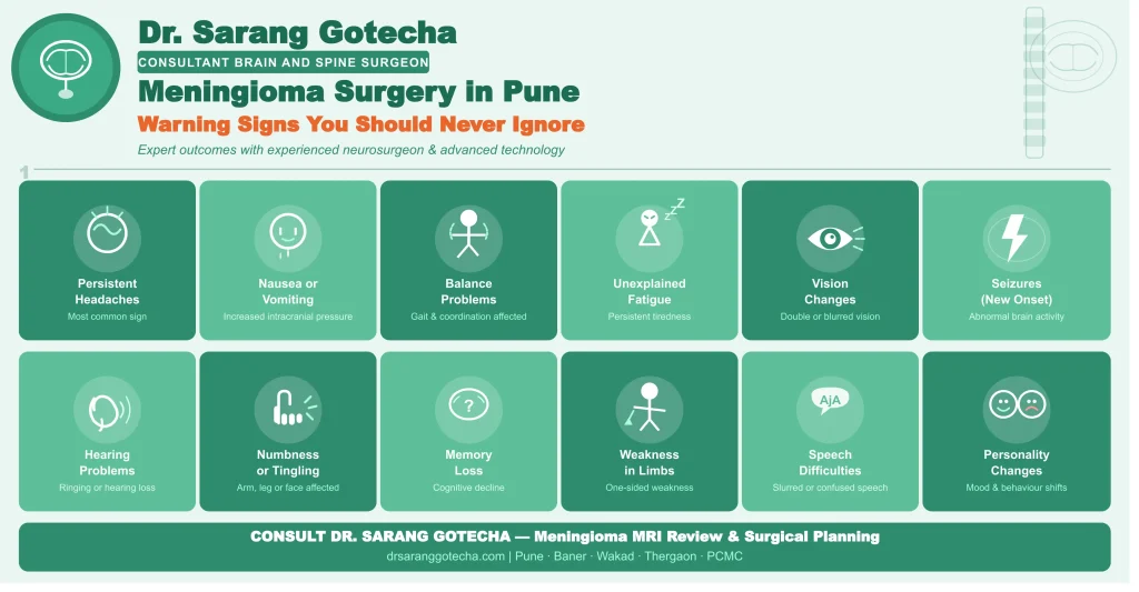
| Symptomatic — headache, seizure, deficit | Surgery recommended | Symptoms indicate mass effect |
| Compressing optic nerve/chiasm | Urgent surgery | Vision loss risk — reversible if treated early |
| Grade 2 or 3 on biopsy | Surgery + adjuvant radiation | Higher recurrence — aggressive management warranted |
| Rapid growth on serial MRI | Surgery — regardless of symptoms | Growing meningioma will become symptomatic |
The Simpson Grading System: Why Extent of Resection Determines Recurrence
The Simpson Grading system — published in 1957 and still the standard — classifies meningioma resection into five grades based on completeness of removal. It directly predicts long-term recurrence rates.
| Simpson Grade | What Was Removed | 10-Year Recurrence Rate |
| Grade I | Complete removal including dural attachment + abnormal bone | Under 9% |
| Grade II | Complete removal + dural coagulation (no excision) | 10 to 15% |
| Grade III | Complete removal — dural attachment not addressed | 20 to 30% |
| Grade IV | Partial removal — subtotal resection | 30 to 40%+ |
| Grade V | Biopsy only | Tumor continues to grow |
For convexity meningiomas, Simpson Grade I resection is achievable in over 90% of cases — and this is the grade associated with the lowest recurrence. For skull base meningiomas involving the cavernous sinus or encasing major vessels, Simpson Grade I is often not achievable without unacceptable cranial nerve morbidity. In these cases, planned Grade III or IV resection followed by stereotactic radiosurgery (Gamma Knife) for residual tumor is the safer, functionally superior strategy.
Surgical Approaches for Meningioma in Pune
Convexity Meningioma
Convexity meningiomas arise from the dura of the brain’s outer surface and are the most surgically accessible type. A craniotomy centred over the tumor with neuronavigation guidance provides excellent exposure. The dural attachment is coagulated and excised (Simpson Grade I). Blood supply from feeding meningeal vessels is interrupted before the tumor is removed. Most patients are discharged in 5 to 7 days.
Parasagittal and Falcine Meningioma
These tumors arise adjacent to the superior sagittal sinus and falx cerebri. When they invade the sinus — particularly the posterior two-thirds which drains critical venous blood — complete resection risks life-threatening venous haemorrhage. The extent of sinus invasion on MRI venography determines the surgical strategy. For patent sinus invasion, planned subtotal resection plus radiosurgery is safer than attempting sinus resection.
Sphenoid Wing and Skull Base Meningioma
Sphenoid wing meningiomas range from easily resectable lateral sphenoid lesions to complex en-plaque tumors wrapping around the carotid artery and invading the cavernous sinus. The surgical approach is the pterional or orbitozygomatic craniotomy. Cavernous sinus involvement is the single most important factor limiting GTR — the cavernous sinus contains the internal carotid artery and cranial nerves III, IV, V1, V2 and VI, none of which can be sacrificed.
Pre-Operative Preparation and Post-Operative Care for Meningioma Surgery
Pre-operative workup includes gadolinium-enhanced MRI (within 3 months), CT angiography for vascular anatomy in skull base tumors, MRI venography for parasagittal lesions invading the superior sagittal sinus, formal visual field testing for tumors near the optic apparatus, and anaesthesia fitness assessment. For hypervascular meningiomas, pre-operative embolisation reduces intraoperative blood loss significantly.
Post-operatively, most meningioma patients are monitored in the neurosurgical ICU for 24 to 48 hours before transfer to the ward. Anti-epileptic medications are prescribed for patients with peri-tumoral oedema or seizure at presentation. Corticosteroids (dexamethasone) are tapered over 3 to 5 days to manage post-operative oedema. Physiotherapy begins on Day 2 to 3.
| Recovery Milestone | Timeline |
| Neurosurgical ICU stay | 1 to 2 days (standard); 3 to 5 days (skull base) |
| Ward stay post-ICU | 3 to 5 days |
| Steroid taper completion | Day 5 to 7 post-surgery |
| Scalp suture/staple removal | Day 10 to 14 |
| Return to desk work | 6 to 8 weeks |
| Driving | 8 to 10 weeks (surgeon clearance) |
| Return to full physical activity | 3 to 4 months |
| First post-op MRI | 6 to 8 weeks |
| Long-term surveillance MRI | Annual for 5 years then every 2 years |
Meningioma Surgery Cost in Pune 2026
| Tumor Location | Cost Range (INR) | Hospital Stay | Notes |
| Convexity meningioma | 2,50,000 to 4,50,000 | 5 to 7 days | Most accessible — best outcomes |
| Parasagittal / falcine | 3,00,000 to 5,50,000 | 5 to 8 days | Sinus involvement affects approach |
| Sphenoid wing (lateral) | 3,50,000 to 6,00,000 | 6 to 8 days | Pterional craniotomy |
| Sphenoid wing (medial/cavernous sinus) | 5,00,000 to 8,00,000 | 7 to 12 days | Skull base approach, IONM mandatory |
| Olfactory groove | 3,50,000 to 6,00,000 | 5 to 8 days | Bifrontal craniotomy |
| Petroclival | 6,00,000 to 10,00,000 | 8 to 14 days | Most complex — transpetrosal approach |
| Spinal meningioma | 2,50,000 to 4,50,000 | 5 to 7 days | Laminectomy + microsurgery |
Meningioma Treatment in Pune and PCMC
A 52-year-old woman from Wakad was found to have a 4.2 cm right convexity meningioma on MRI performed for progressive headaches over 6 months. Formal visual field testing was normal. Pre-operative embolisation was performed for a highly vascular tumor. Pterional craniotomy with neuronavigation achieved Simpson Grade I resection — complete removal including the dural attachment. She was discharged on Day 6 with no neurological deficit. Post-operative MRI at 6 weeks confirmed no residual tumor. She returned to her teaching role at 7 weeks.
Dr. Sarang Gotecha performs meningioma surgery at hospitals in the Baner-Wakad corridor with full neuronavigation, IONM and ICU support. For meningioma consultation, MRI review and surgical planning, book at drsaranggotecha.com.
Frequently Asked Questions
Q: What is the success rate of meningioma surgery in Pune?
A: For convexity meningiomas — the most common and accessible type — gross total resection is achieved in 85 to 95% of cases at experienced neurosurgical centres in Pune. The 10-year recurrence rate after Simpson Grade I complete resection of WHO Grade 1 meningioma is under 9%. For skull base meningiomas involving the cavernous sinus, subtotal resection rates are higher — planned residual plus Gamma Knife radiosurgery is the standard approach.
Q: Does every meningioma in Pune need surgery?
A: No. Small asymptomatic meningiomas (under 3 cm) in adults over 65 years can be safely observed with MRI every 6 to 12 months. Surgery is recommended when: the tumor is symptomatic (causing headache, seizure or neurological deficit), it is compressing the optic nerve or chiasm, it is over 3 cm, or it shows significant growth on serial imaging. The decision is individualised based on tumor characteristics and patient health.
Q: What is the cost of meningioma surgery in Pune in 2026?
A: Meningioma surgery cost in Pune in 2026 ranges from INR 2,50,000 to INR 4,50,000 for accessible convexity tumors to INR 6,00,000 to INR 10,00,000 for complex skull base or petroclival meningiomas. Costs include surgeon, anaesthesia, neuronavigation, IONM where required, ICU stay, ward stay and post-operative medications. Written itemised quotes should always be obtained before surgery.
Q: What is the Simpson Grade and why does it matter for meningioma?
A: The Simpson Grading system classifies the completeness of meningioma resection at surgery: Grade I (complete removal including dural attachment and abnormal bone) has under 9% recurrence at 10 years. Grade IV (partial removal) has over 40% recurrence. It is the most important predictor of long-term outcome after meningioma surgery. Patients should ask their surgeon what Simpson Grade they plan to achieve and why.
Q: How long is recovery after meningioma surgery in Pune?
A: Hospital stay after meningioma surgery is 5 to 8 days for standard craniotomy and 7 to 14 days for complex skull base approaches. Return to desk work is 6 to 8 weeks. Return to full physical activity is 3 to 4 months. The first post-operative MRI is performed at 6 to 8 weeks to confirm resection completeness. Long-term surveillance MRI is recommended annually for 5 years then every 2 years.
Q: Is meningioma surgery covered under health insurance in India?
A: Yes. Meningioma surgery including craniotomy, skull base approaches and post-operative ICU care is covered under most comprehensive private health insurance policies in India. Pre-authorisation from your TPA is required before surgery. Provide gadolinium-enhanced MRI reports and the surgeon’s operative plan. Ayushman Bharat PM-JAY covers standard craniotomy for benign brain tumors at empanelled hospitals.
Meningioma surgery in Pune offers patients excellent outcomes when performed by an experienced neurosurgeon with appropriate technology. The majority of convexity meningiomas are cured with a single operation. For complex skull base tumors, a planned strategy combining maximal safe resection with adjuvant radiosurgery provides the best functional and oncological outcome.
For meningioma consultation, MRI review and surgical planning with Dr. Sarang Gotecha, book at drsaranggotecha.com. Serving patients across Pune, Baner, Wakad, Thergaon and PCMC.
Book Your Consultation With Dr. Sarang Gotecha

Dr. Sarang Gotecha
Dr. Sarang Gotecha is a leading brain & spine surgeon in Pune, offering advanced care for complex neurological and spinal conditions. With strong academic credentials (MBBS, MS, MCh Neurosurgery) and years of surgical experience, he is committed to delivering precise, safe, and patient-focused treatments.
- Expert in brain tumor, spine & neuroendoscopic surgeries
- Specialized in minimally invasive & skull base surgeries
- Follows an ethical and patient-centric approach
- Available at clinics in Baner, Wakad, and Thergaon (Pune)